
Developing microwave-induced thermoacoustic tomography: System,application,and reconstruction
2024-01-17 03:06:08ShungLiLiuWnTingPengXioZhngZhuJinSongXinShngHuZhngZhiQinZho
Shung-Li Liu,Wn-Ting Peng,Xio-Zhng Zhu,Jin Song,Xin Shng,Hu Zhng,Zhi-Qin Zho,*
a School of Information Engineering,Southwest University of Science and Technology,Mianyang,621010,China
b School of Electronic Science and Engineering,University of Electronic Science and Technology of China,Chengdu,611731,China
c Yunnan Key Laboratory of Computer Technologies Application,Kunming University of Science and Technology,Kunming,650500,China
Keywords:Application Microwave-induced thermoacoustic tomography(MITAT)Reconstruction System
ABSTRACT Amicrowave-induced thermoacoustic imaging (MITAT) system is a non-destructive physical medical imaging method that combines the advantages of the high contrast of microwave imaging and the high resolution of ultrasound imaging.It uses the microwave as the excitation source and ultrasound as the information carrier.When different kinds of biological tissue absorb electromagnetic energy,it results in localized temperature rises.The thermal expansion will induce ultrasonic signals (i.e.,thermoacoustic signals),known as the thermoacoustic effect.The microwave absorption image of the sample can be reconstructed by algorithm processing.The MITAT contrast depends on different dielectric parameters of different kinds of tissue.We introduce the developed system and its application.In addition,the challenges and prospects of MITAT for further development are discussed.
1 Introduction
Medical imaging plays a crucial role in the clinical diagnostic field.Among the existing medical imaging methods,microwave thermoacoustic imaging techniques can be summarized in Fig.1.First,the pulsed microwave is used as an excitation source to radiate biological tissue.Second,biological tissue absorbs microwave energy and then expands,producing a mechanical wave (or a thermoacoustic signal),and the thermoacoustic signal radiates outward.After receiving the signal,a probe amplifies the signal.The acquisition card collects the signal.Finally,an image is reconstructed using a relevant algorithm.According to the type of electromagnetic pulses,microwave thermoacoustic imaging is generally divided into photoacoustic imaging (PAI) and microwave-induced thermoacoustic tomography imaging (MITAT).Both can achieve high-resolution and high-contrast imaging.Yet,owing to the high frequency of photoacoustic excitation sources in PAI,the PAI-related methods usually focus on superficial tissue [1].On the other hand,the MITAT technology has potential advantages in detecting deep tissue and has attracted the attention of many researchers [2].

Fig.1.Basic schematic diagram of thermoacoustic imaging: (a) microwave pulses exciting biological tissue,(b) thermoelastic expansion of biological tissue producing ultrasonic waves,and (c) distribution of microwave energy absorption density directly reconstructed by the thermoacoustic image.
In the 1980s,the application of the thermoacoustic effect in imaging began to be studied.Bowen [3]pioneered its application in soft tissue imaging.In 1984,Guo et al.[4] studied the generation mechanism of microwave thermoacoustic signals in combination with thermodynamic processes,explored the discontinuity of thermodynamic variables and electromagnetic energy absorption in biological tissue,and gave the general formula of MITAT.In 1999,Kruger et al.[5,6] studied thermoacoustic breast imaging at 434 MHz.In 2000,Wang’s group [7,8] further improved and developed the MITAT system,and built an imaging system with the scanning mode.They analyzed the system’s thermoacoustic signal,image resolution,and contrast.In 2002,Wang’s group [9–11] studied and analyzed different imaging array modes (including planar array,cylindrical array,and spherical array) in the time-frequency domain.Subsequently,researchers realized the superiority of MITAT and devoted themselves to enhancing the performance of the MITAT system.At present,the main MITAT research groups include Patch et al.[12–15],Ntziachristos’s group [16,17],Jiang’s group [18–20],Wang et al.[21],Nan et al.[22],Zheng’s group [23–25],Xing’s group [26,27],Wang et al.[28,29],and Song [30].The MITAT system and its application in the biological field have made incredible breakthroughs.
The excitation source is the core component of the thermoacoustic imaging system.Table 1 briefly summarizes the key parameters of the microwave excitation source currently used by each group,including frequency,pulse width,peak power (voltage),and repetition frequency.These research groups may focus on various aspects of the microwave thermoacoustic imaging technology.Their research may involve theoretical analyses of factors affecting image quality,such as the influence of limited probe bandwidth and aperture,limited angle detection,uneven sound velocity,uneven field distribution,and pulse width.Alternatively,they may prioritize the improvement of imaging systems for faster and better imaging,including multi-modal imaging,large field of three-dimensional (3D) view imaging,fast or real-time imaging of array probes,ultrashort microwave pulse radiation,uniform field distribution antenna design,microwave energy focusing,negative acoustic lens,and adaptive surface probes.

Table 1 Comparison of microwave excitation source parameters in each study.
Moreover,the research groups may concentrate on specific applications such as breast disease,foreign body detection,kidney imaging,brain tissue imaging,blood vessel imaging,and thyroid imaging.Some may emphasize a comprehensive approach,covering theory,system algorithms,and applications.Additionally,they may study thermoacoustic molecular imaging using contrast agents,thermoacoustic spectral imaging,thermoacoustic temperature monitoring,continuous microwave-induced thermoacoustic imaging,and circuit modeling of thermoacoustic effects.The diverse studies collectively contribute to advancing the microwave thermoacoustic imaging technology.
Since 2005,Zhao’s team from University of Electronic Science and Technology of China has explored the MITAT technology,including imaging systems,application fields,and reconstructed algorithms.The related work of the team in the MITAT field is presented.First,the development of the MITAT system is described.Then,various kinds of biological tissue are examined with the developed system.Finally,the challenges of microwave thermoacoustic imaging are discussed.
2 Microwave-induced thermoacoustic imaging system
The main technological advantage of the MITAT system comes from the multi-physical field conversion of the electromagnetic field combined with the ultrasonic field so that the microwave-induced thermoacoustic image can have the edges of the high contrast of microwave imaging and high resolution of ultrasonic imaging.The system mainly includes the microwave excitation module,thermoacoustic signal-receiving device,and image processing equipment.Each is detailed next.
2.1 Microwave excitation module
The microwave pulse source usually consists of the magnetic-controlled modulation mode,high voltage discharge mode,and modulation continuous mode.The magnetic-controlled modulation mode enhances the intensity of the thermoacoustic signal and reduces the noise.Thus,the signal-to-noise (SNR) ratio is improved.The mode is the primary one for the excitation source.As shown in Fig.2 (a),an electron emitter,power supply,magnetron,and waveguide typically make up the microwave generator of this mode.The electron emitter in the magnetron can emit electrons,and electrons are accelerated and produce stable electron oscillation.The waveguide modulates the electron oscillation in the microwave,and the waveguide mode of the microwave is radiational.This mode allows the sample to be better radiated by concentrated energy.Thus,the thermoacoustic signal can be stimulated,and the SNR ratio can be improved,which is of great significance for enhancing imaging quality.
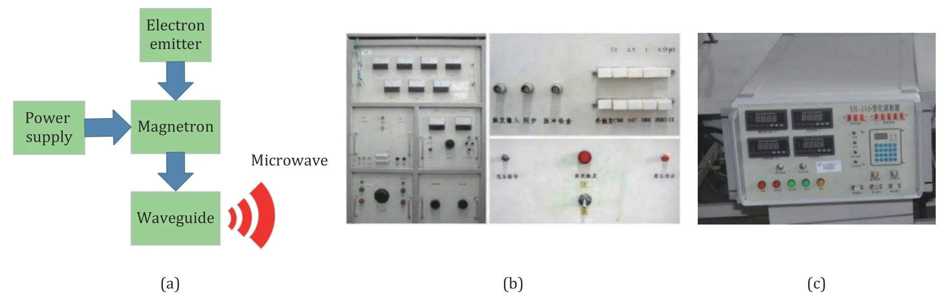
Fig.2.Microwave sources: (a) schematic diagram of the magnetron modulation mode,(b) large source,and (c) small source.
The microwave pulse excitation part was improved to make the microwave pulse source smaller [31].The solid-state modulator is used to replace the linear modulator,thereby reducing the cost and the quality.In the solid-state modulator,the volume of the pulse transformer with a high conversion ratio (1∶20) can be reduced by about 4/5 under the same voltage and power conditions.Figs.2 (b) and (c) show large and small microwave pulse sources,respectively.Studies have shown that high-power microwave sources with the nanosecond pulse width and megawatt power can excite broadband thermoacoustic signals to achieve sub-millimeter resolution.
2.2 Thermoacoustic signal-receiving device
The receiving device of the thermoacoustic signal is usually a single sensor or a sensor array,and the detection method varies according to the layout of the sensor array.In tomography,the image is usually two-dimensional (2D),which requires the sensor layout to be a linear array or 2D around,not just a point.The commonly used receiver sensor layout is shown in Fig.3.The array layout uses a single sensor for 2D rotation (some experimental systems use a fixed ultrasonic sensor to rotate the sample to be tested by 2D rotation) or Z-shaped movement.Perhaps the array layout directly uses the sensor array.The sensor array is usually divided into circular and linear arrays [32].
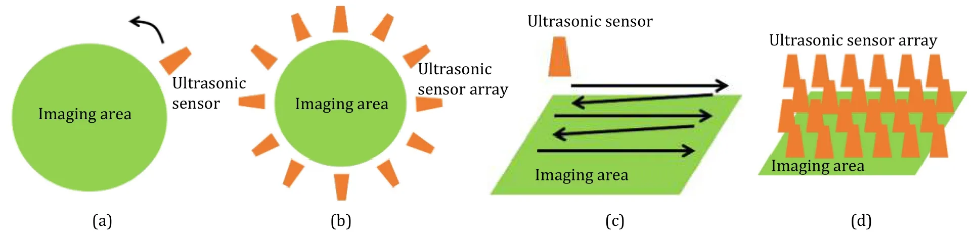
Fig.3.Sensor layout of a common receiving device: (a) single sensor 2D rotation,(b) sensor array 2D around,(c) single sensor linear scan,and (d) linear sensor array.
The sensor layout of the receiving device has gone through the following stages of development.First,the 2D stepping scanning device of Arrick Robotics Company in the USA was modified.A belt drive became a screw drive [33].Fig.4 (a) shows the screw drive scanning platform.Second,a rotating platform was designed to miniaturize the system,as shown in Fig.4 (b).The transmission platform uses a stepper motor to drive an annular bearing device,and a bracket is installed on the bearing device.The probe and the radiation antenna are installed on the bracket,and the computer controls the stepper motor while collecting the generated thermoacoustic signal.Finally,an array sensor bowl [34] was designed to improve patient comfort.Fig.4 (c) shows the designed coupled liquid container with an ultrasonic sensor.Table 2 compares the receiving device of the thermoacoustic signal.
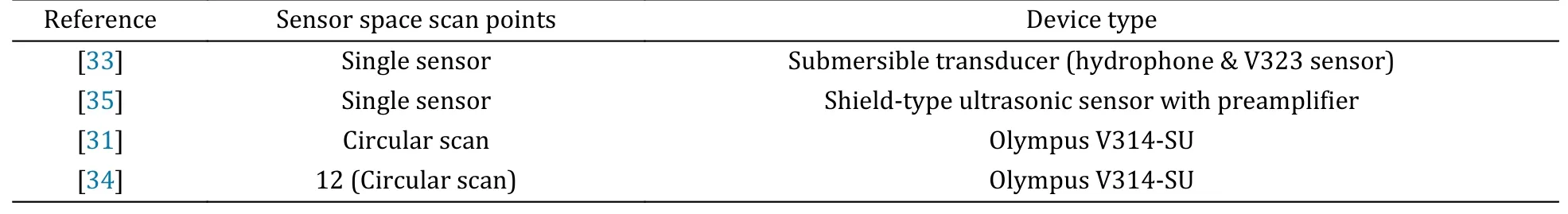
Table 2 Comparison of thermoacoustic signal-receiving devices in MITAT systems.
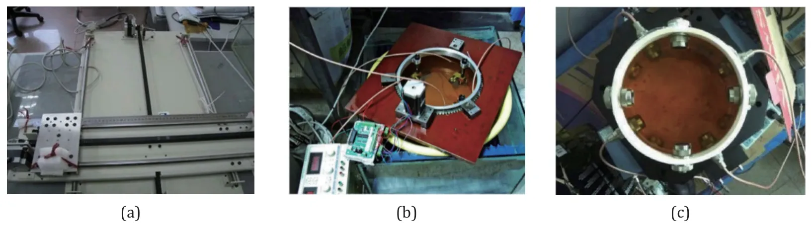
Fig.4.Thermoacoustic signal-receiving device: (a) screw drive,(b) single transducer scanning,and (c) transducer array scanning in Ref.[34].Reproduced with permission [34].
The criteria for selecting ultrasonic probes exist for a miniaturized microwave thermoacoustic imaging system.First,the frequency response of the ultrasonic probe should be within the frequency range of the microwave-induced thermoacoustic signal.Second,for system miniaturization,the system should be highly integrated.Thus,the ultrasound probe is required to have a smaller size.A typical probe’s cross-sectional size should be controlled at about 20 cm.Third,the coupling liquid is needed in the detection to avoid the attenuation of ultrasound,so the probe must be able to work normally under water or in liquid.Fourth,the ultrasonic probe will work properly in a strong electromagnetic environment in this system.Therefore,the probe must have the anti-interference ability to microwave pulses.Ultimately,the team chose the Olympus V314-SU as the probe used in the system experiments,with a center frequency of 1 MHz.
2.3 Image processing equipment
The image data processing is the most crucial part of the imaging process,which relates to whether the imaging results can truly and completely reconstruct the sample structure,size,and location information.As data processing equipment,the computer plays an indispensable role.We integrated vital components such as the image display,system host,and system controller into the standard 19-inch cabinet,which not only improves the stability of the system but also enhances its maintainability [36],as shown in Fig.5.In the system design,the patient is assumed to lie prone on the examination bed,and the breast to be detected is immersed in the ultrasonic coupling liquid of the container through the examination hole.The doctor sets up operations in front of the cabinet through the human-computer interaction interface of the system and controls the scanning platform to perform spatial scanning to obtain thermoacoustic signals.With a scanning step angle of 1.8 degrees,the scanning time is about 2 minutes.After the scanning is completed,the thermoacoustic image reconstruction can be directly performed in the human-computer interaction interface,and the detection results can be observed.
Fig.5.MITAT system diagram.
The image reconstruction can be divided into direct and indirect reconstructions.The direct reconstruction uses the sensor and spatial information of the imaging region for direct imaging without mathematical models.The indirect reconstruction requires appropriate mathematical tools to model the physical imaging process and invert the image according to the model.Therefore,the process of the indirect reconstruction includes mathematical modeling and the image reconstruction.At present,most computed tomography images are indirect reconstructions.
2.3.1 Back projection algorithm
The back projection (BP) algorithm ignores the non-uniformity of biological tissue and the scattering and diffraction of broadband ultrasonic waves in biological tissue.The algorithm reverses the time of the received microwave thermoacoustic signal and then projects it back to the imaging area.Its imaging speed is fast,but the imaging quality is poor.The reconstruction expression of the BP algorithm is shown by
whereΩis a sector contained by a circular arc in two dimensions and a cone corresponding to a spherical surface element in three dimensions,Ploss(r′) is the energy density absorbed by biological tissue per unit volume per unit time,rrepresents the position of the sound pressure field point,r′represents the position of the sound pressure source point,trepresents the time when the sound pressure reachesr,andcis the speed of sound in tissue which is related to tissue properties.Equation (1) includes the thermoacoustic signal termp(r,t) and the first-order partial differential of the thermoacoustic signal with respect to time.The BP algorithm containing only the thermoacoustic signal term is called the delay and sum (DAS) algorithm.The BP algorithm containing these two terms is called the filtered back projection (FBP) algorithm [37].Compared with DAS,FBP introduces high-pass filtering characteristics,but there is no essential difference in principle between the two algorithms.
The BP and DAS algorithms are likely to be understood and implemented.For all that,these two methods neglect the influence of microwaves.Second,these two methods assume that the speed of sound in biological tissue is invariable.That may be inconsistent with the actual situation.Third,they cannot derive the functional information of biological tissue.
2.3.2 Time reversal mirror algorithm
The time reversal mirror (TRM) algorithm is an accurate mathematical model.Fig.6 shows the principle of the TRM algorithm [38].Time reversal is the physical mechanism that the TRM algorithm follows.The TRM method needs to consider the physical process of sound wave propagation and solve the wave equation through appropriate boundary conditions and numerical methods.The TRM algorithm has better inversion accuracy but more computation than the BP algorithm.

Fig.6.TRM principle: (a) forward wave propagation generated by a thermoacoustic source and received by a sensor array in the time domain and (b) received signals with a time reversal sequence re-emitted into the media and refocused at the original position.
2.3.3 Compressed sensing algorithm
Although the BP and TRM algorithms are robust and easy to implement,they require highly intensive spatial sampling of the acoustic signal and high-quality 3D sample images to be obtained.When MITAT detects breast cancer,large spatial sampling points are needed,inevitably leading to long scanning time.A new method that combines MITAT with compressed sensing (CS) solves the problems faced [39].Fig.7 (a)shows a graphical representation of the process in which the MITA signal received by the sensor array is represented by an overcomplete dictionary based on the anatomical breast model.The total response ofNtargets measured by the sensor array can be written asB.The imaging process aims to recover the information vectorbfromB,as shown in Fig.7 (b).Compared with the traditional time-domain BP and TRM methods,the sparse sampling-based compressed sensing thermoacoustic imaging (CS-MITAT) method can significantly reduce the data acquisition time and improve imaging quality.
Fig.7.MITAT procedures represented by dictionaries in Ref.[39]: (a) MITAT process expressed by the over-complete dictionary and (b) spatial compressive sensing procedure.Reproduced with permission [39].
The existing CS-MITAT algorithm has two main problems.One is that the high-resolution image reconstruction time is long.The other is that the establishment time of the thermoacoustic dictionary is long,and the memory occupation is large.To solve the first problem,we proposed a multilayer dictionary compressed sensing thermoacoustic imaging (HDCS-MITAT) method to reduce the computational cost of high-resolution reconstruction [40].Different dictionaries have different spatial resolution.In image reconstruction,the low-resolution dictionary estimates the target region.Then,the high-resolution dictionary is used to image the estimated region to reduce the reconstruction calculation.Fig.8 shows the schematic diagram of HDCS-MITAT.Table 3 shows the reconstruction time of HDCS-MITAT and CS-MITAT under different numbers of sensorsK.The results show that compared with CS-MITAT,HDCS-MITAT has less computation time.To solve the second problem,we studied a fast method of dictionary construction by simplifying the solution of the wave equation,which can significantly reduce the time and memory consumption of thermoacoustic dictionary construction.The thermoacoustic dictionary matrix is compressed into a sparse matrix using the characteristics of the sparse time domain of the thermal sound generated by a fine grid source [41].Thus,fast build time and memory savings are achieved.

Table 3 Time comparisons of HDCS-MITAT and CS-MITAT in Ref.[40].Reproduced with permission [40].
Fig.8.Schematic diagram of the HDCS-MITAT algorithm in Ref.[40].Reproduced with permission [40].
2.3.4 Frequency-domain imaging algorithm
The frequency-domain imaging algorithm is somewhat similar to the FBP imaging algorithm mentioned earlier,except they deal with the same problem in different spaces [42].The frequency domain imaging algorithm processes data to improve the calculation efficiency in the frequency domain.It is observed that the derivation process from (2) to (3),compared with the FBP algorithm,the frequency-domain imaging algorithm does not do any approximate processing.Therefore,theoretically,the frequency-domain imaging algorithm has better imaging quality.
The microwave thermoacoustic wave equation can be described by
wherevs=is the propagation velocity of ultrasound,p(r,t) is the sound pressure,H(r′,t) is the absorbed microwave energy distribution at any positionrand at any timet,Cis the specific heat capacity,andβis the isobaric volume expansion coefficient.
Using the Fourier transform and Green’s theorem,(2) can be transformed into:
whereΦ(u,v,w) is the distribution of the microwave energy deposition,ρis the density of muscle tissue,Pd(x,y,t) is the signal from the detector,is the temporal and spatial Fourier transform ofPd(x,y,t),I0is a scaling factor proportional to the incident radiation intensity,ηis the shape of the irradiating pulse,denotes the Fourier transforms ofη,R(x,y) describes the size of the detector,andis the spatial Fourier transform ofR(x,y).Map the (u,v,k) space into the (u,v,w) space by the relationw=.Although the frequency domain imaging algorithm has advantages theoretically in the practical application,it needs to consider the factors including the computational complexity,interpolation error,application range,and real-time.These factors may limit its application.
3 System application
Biological tissue with different dielectric parameters has different microwave absorption characteristics,so the MITAT technology can image biological tissue with a high contrast and high resolution and visualize deep pathological tissue.Currently,MITAT has potential applications in biomedical imaging fields including breast cancer diagnosis,prostate evaluation,joint detection and evaluation,brain imaging,thermoacoustic endoscopy (TAE) examination,and angiography.Here,we delve into the examination of the contrast and resolution of our imaging system,with a particular focus on system’s application in various types of tissues.These include,but are not limited to,breast tissue,brain tissue,and liver tissue.
3.1 Imaging contrast
The contrast of microwave thermoacoustic imaging depends on the dielectric parameters of biological tissue.When the microwave thermoacoustic wave passes through the biological tissue,the dielectric constant determines the speed and direction of the electromagnetic wave propagation inside the tissue.It causes the reflection and refraction of the electromagnetic wave.Different types of tissue differ in permittivity.The differences show up as different reflected and scattered signals in imaging,ultimately affecting the image contrast.After comparing agar with gelatin,an agar-water mixture was imitated as the main component to simulate biological tissue [31].The effect of the final prepared phantom is shown in Fig.9.The electrical and acoustic parameters of the phantom can be adjusted,and the imitations can undergo arbitrary plasticity based on the shape of the mold in terms of appearance.

Fig.9.Biological tissue phantom sample photos.
The imaging contrast sources of MITAT can be generally divided into endogenous and exogenous.Here,polar molecules and charged ions are the two main sources of endogenous contrast agents.Exogenous contrast agents can also improve the contrast of thermoacoustic images.A major research interest in MITAT is the development of exogenous contrast agents with biocompatibility,such as targeted NaCl nanodroplets.Exogenous contrast agents with biocompatibility can track cancer growth without interfering with the internal environment,thus having a great application value in early cancer detection.
Carbon nanotubes are used as an exogenous contrast agent for thermoacoustic imaging.Figs.10 (a) and(b) compare dielectric parameters of cancerous tissue containing carbon nanotubes.According to the results of using carbon nanotubes in microwave imaging of breast cancer,carbon nanotubes,with their high conductivity,can effectively improve the contrast of node parameters.Fig.10 (c) shows the peak-to-peak values of the thermoacoustic signals of four kinds of tissue received by the ultrasonic sensor at different receiving angles (100 groups).The results show that carbon nanotubes,as a contrast agent for thermoacoustic imaging,can enhance the absorption of microwave energy by increasing the dielectric parameters of tissue,thus improving the image contrast [43].Then,the conductivity and speed of sound of the simulated material were studied.Figs.10 (d) and (e) show the conductivity and sound velocity of the simulated material with different mass ratios of contrast agents.The results show that the electrical conductivity and sound velocity of the phantom material increase with the increase in the content of carbon nanotubes [44].Furthermore,the thermoacoustic response of carbon nanotube phantoms with different contents was studied.TheA,B,C,andDmarked in Fig.10 (f) are the carbon nanotubes with mass ratios of 1.00%,0.50%,and 0.22%,respectively,and the carbon nanotube-free phantom samples.The results show that carbon nanotubes as a contrast agent can effectively increase the contrast between the target and the surrounding tissue in the MITAT technology.

Fig.10.Effect of carbon nanotubes (with the abbreviation CNTs in the figure) on the dielectric parameters of materials in Ref.[44]: (a) relative dielectric constant,(b) electrical conductivity,(c) thermoacoustic response statistics of various substances,including fat,gland,tumor,and tumor-containing contrast agents,(d) electrical conductivity of phantom materials measured at different mass ratios of contrast agents,(e) sound velocity values determined for phantom materials with varying mass ratios of contrast agents,and (f) thermoacoustic images obtained for carbon nanotubes with different contents.Reproduced with permission [44].
As a material with great application potential in the biomedical field,carbon nanotubes have achieved many research results in cancer treatment,gene therapy,and drug-targeted delivery [45].Among them,the toxicity study of carbon nanotubes is the basic research of its application in biomedicine.It is the foundation of the application of carbon nanotubes in biomedicine.According to Ref.[46],the content of carbon nanotubes is a vital factor affecting their toxicity.Therefore,based on the consideration of application safety,the phantom sample with the lowest carbon nanotube content (0.22%) was used in the thermoacoustic imaging experiment.In the imaging experiment of different sample shapes,the samples containing carbon nanotubes showed a stronger thermoacoustic response than those without carbon nanotubes.
3.2 Image resolution
From the point of view of image processing,a thermoacoustic imaging system can be seen as a cascade of several systems.Fig.11 is the system cascading diagram.According to the system cascading theory,sensors and amplifiers will impact the spectrum of the final detected piezoelectric signal,thus affecting the final resolution.The sensor’s bandwidth should include the frequency spectrum of the ultrasonic signal to achieve high resolution.The amplifier’s bandwidth should be large enough to contain the spectrum of the entire ultrasonic signal so that the final system resolution will not decrease due to improper hardware selection.Additionally,the resolution of the system is also affected by the spatial sampling spacing of the sensor receiving array and the uniformity of the microwave pulse radiation.

Fig.11.System cascading diagram.
In the current MITAT technology,the microwave pulse width is generally about 1 μs,and the peak power is in the order of kW.To increase the heat accumulation in biological tissue,we increase the width of the microwave pulse,thereby increasing SNR of the thermoacoustic signal.However,the research results show that the width of the thermoacoustic signal is proportional to that of the microwave pulse.The microwave pulse as the excitation source must be as short as possible to reach high-resolution thermoacoustic images [47].The different requirements of the microwave pulse width form a contradiction between SNR of the thermoacoustic signal and the image resolution.To improve the resolution of the thermoacoustic image without affecting SNR of the thermoacoustic signal excited by the biological tissue,Song [30] studied the high-power short-pulse excitation thermoacoustic imaging system.Fig.12 is a schematic diagram of the high-power short-pulse thermoacoustic experiment.The experimental results obtained the thermoacoustic signal bandwidth superior to the current thermoacoustic imaging system.The high-power short-pulse excitation thermoacoustic imaging system provides the feasibility for further improving the resolution of the thermoacoustic image.Subsequently,the thermoacoustic effect excited by a short-pulse microwave source was explored,and the influence of two different microwave pulses on the imaging quality was studied.The imaging results in Fig.13 show that the thermoacoustic signal excited by a high-power short pulse source boosts the image resolution [48].
Fig.12.High power short pulse thermoacoustic experiment: (a) experimental diagram,(b) time-domain waveform of the thermoacoustic signal,and (c) thermoacoustic signal spectrum.
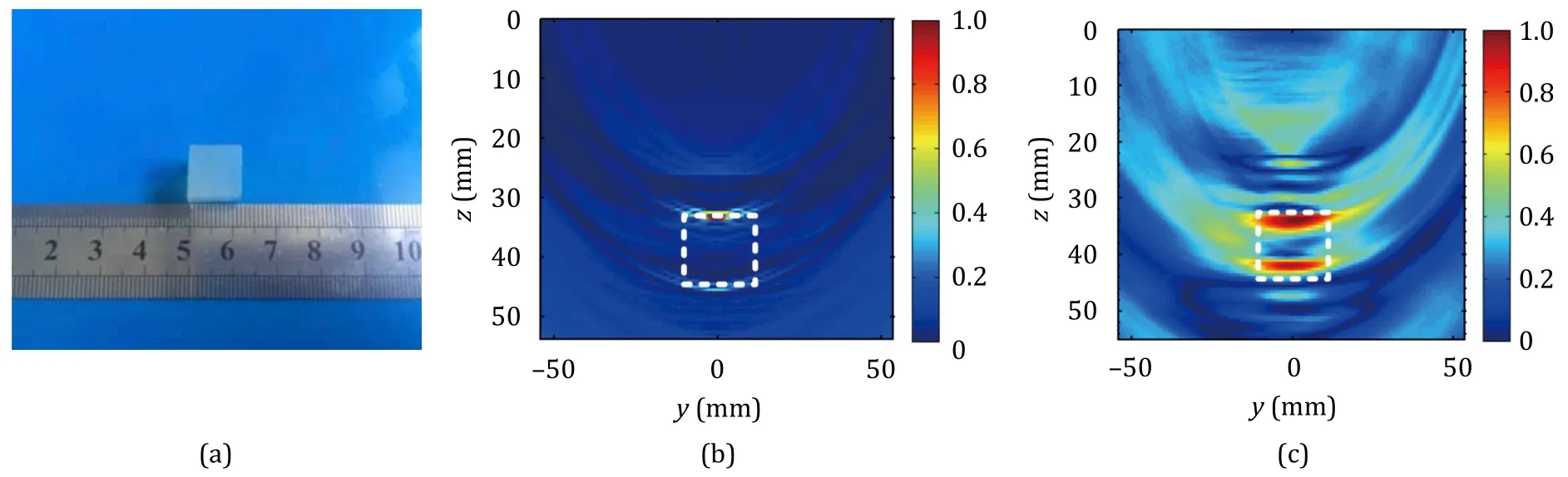
Fig.13.Thermoacoustic signal by the same phantom excited by two microwave pulse sources in Ref.[48]: (a) agar phantom,(b) thermoacoustic signal excited by 10 ns microwave pulse source,and (c) thermoacoustic signal excited by 500 ns microwave pulse source.Reproduced with permission [48].
3.3 Microwave thermoacoustic imaging of breast tumor
Breast cancer is one of the most common cancers in women,and its incidence and mortality are rising rapidly.The existing research shows that the early detection and treatment of breast cancer are of great significance in improving the survival rate and cure rate of breast cancer patients.The difference in electrical conductivity between malignant breast tissue and normal breast tissue is 6∶1,and the difference in electrical conductivity between malignant breast tissue and normal fatty breast tissue is 10∶1 [49].Therefore,it is possible to detect breast tumors by the MITAT technique.
Song cooperated with West China Hospital of Sichuan University,and thein vitroexperiments were conducted on real-stage T1 and T2 breast cancer tissue,invasive ductal carcinoma tissue,and fibroma.In the ultrasonic image shown in Fig.14 (a),it is challenging to distinguish two samples according to their image intensity.Figs.14 (b) and (c) show that the thermoacoustic technology can distinguish between normal breast tissue and cancer tissue,and the contrast of breast cancer in different periods is also different [50].The MITAT system provides a sufficient contrast between normal tissue and tumor tissue and can discriminate tumor tissue in diverse clinical phases.
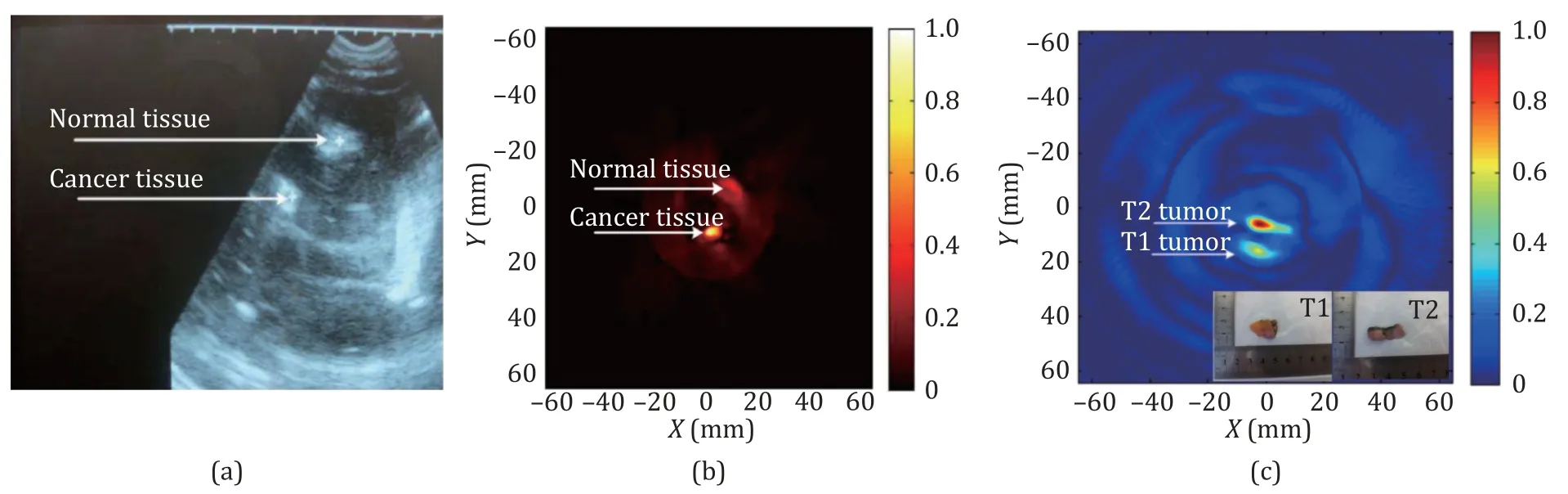
Fig.14.Comparison of ultrasound and thermoacoustic images of real breast tissue and tumor tissue in Ref.[50]:(a) ultrasound image,(b) thermoacoustic image,and (c) thermoacoustic image of T1 (appearance similar to normal adipose tissue,but cancerous) and T2 tumor specimens.Reproduced with permission [50].
Wang et al.conducted microwave thermoacoustic imaging experiments on isolated breast tissue and breast cancer tissue [40].Figs.15 (a)–(c) show images of isolated breast cancer tissue samples and images of imaging experiments,respectively.The HDCS-MITAT imaging results shown in Fig.15 (d) only show the tumor,and the measurement noise and interference are well suppressed.Compared with real tumor targets,HDCS-MITAT imaging results can clearly image most tumor targets,although there is a little loss of detail.Fig.15 (e) shows the image of breast tissue obtained using the block-based CS-MITAT [51].Fig.16 (a) shows the excised breast tissue,with the tumor target enveloped in adipose tissue.Fig.16 (b) is an image reconstructed using the fast dictionary CS.The results show that MITAT can improve the image contrast without losing the main information,which is conducive to target recognition.
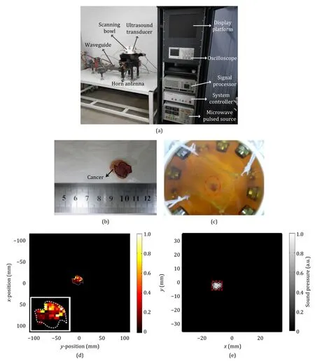
Fig.15.Tumor specimen and its position in the detection container in Ref.[50]: (a) MITAT system,(b) tumor specimen,(c) tumor position,(d) image reconstructed by HDCS,and (e) image reconstructed by block-based CS-MITAT.Reproduced with permission [50].
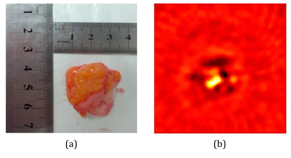
Fig.16.Ex vivo breast tissue image reconstruction with the limited-view scenario in Ref.[41]: (a) picture of ex vivo breast tissue containing a breast tumor (not shown) and(b) CS.Reproduced with permission [41].
3.4 Microwave thermoacoustic brain imaging
The brain is the most structurally and functionally complex tissue in the living body.The difference of microwave absorption and the advantages of high penetration and high resolution of the MITAT technology make MITAT have the advantages of non-ionizing and noninvasive and the potential of real-time high-resolution imaging of living whole brain tissue.In addition,changes in pathological and physiological characteristics of tissue can cause changes in electrical conductivity,which provides a theoretical basis for MITAT to detect diseased tissue.Liu used the MITAT technology to image real pig brains inserted into a saline tube.Fig.17 shows the experimental setup and imaging results.The acoustic velocity self-focusing imaging algorithm based on the Gaussian mixture model (GMM) is used to process it [52].MITAT can clearly distinguish the brain’s central line and reconstruct the circular saline tube targets.The diameters are consistent with reality,and the details of other folds in the brain can also be found.The results of this study indicate that the MITAT technology has great potential in neuroscience research and provides a basis for the clinical application of the MITAT technology in brain imaging.

Fig.17.Experiments of a pig’s brain with a salt tube inside [52]: (a) picture of a pig’s brain with a salt tube inside and(b) image reconstructed based on the proposed method.Reproduced with permission [52].
The microwave thermoacoustic imaging technology provides a new noninvasive brain imaging scheme and a new technical tool for understanding the functional structure of the brain.The technology helps people to explore brain function further.It is expected to become an essential means to study brain function by further developing the real-time brain imaging scheme and overcoming the obstacle of ultrasonic signal interference by the skull.
3.5 Microwave thermoacoustic liver imaging
The liver is an important human organ for metabolizing sugars,proteins,and lipids.With the improvement in living standards,the prevalence of non-alcoholic fatty liver disease caused by obesity or drugs has increased.Fatty liver is mainly due to the accumulation of steatosis in the liver,and the main sources of fat in the liver are food and peripheral fat tissue.Because the early mild fatty liver has no clinical symptoms,the development of severe fatty liver may cause cirrhosis and even liver cancer,so early diagnosis is also crucial for patients with fatty liver.The average conductivity difference between liver and fat can be up to ten times; thanks to microwave penetration,the microwave can stimulate the thermoacoustic effect in the liver to detect fatty liver.
Using the pig liver,lean meat,and fat meat to establish different environments,we carried out an exploratory experiment of microwave thermoacoustic imaging on three samples [32].As shown in Fig.18,the fat and liver are stimulated to produce thermoacoustic effects.However,the liver absorbs more microwave energy due to its higher electrical conductivity compared with fat,thus generating larger thermoacoustic signals.In the reconstructed images,the liver has a higher contrast.Hence,the microwave thermoacoustic images of patients with fatty liver have a darker contrast than those of ordinary people where fat accumulates in the liver or where liver cells undergo degeneration.

Fig.18.Sample and ex vivo tissue imaging: (a) porcine liver sample,(b) pig liver-wrapped lean meat samples,(c) fat-wrapped pig liver samples,(d) TRM imaging for (a),(e) TRM imaging for (b),and (f) TRM imaging for (c).
The above experiments explored the possibility of microwave thermoacoustic signals for imaging and detecting fatty liver usingin vitrobiological tissue.Since the liver is in the abdominal cavity,it needs to be detected with electromagnetic waves with good penetration.Compared with pure ultrasonic imaging,microwave thermoacoustic imaging can combine the high contrast of microwave imaging and the high resolution of ultrasonic imaging,so it has great application prospects in clinical detection.Follow-up studies can be conducted from multiple dimensions,such as microwave excitation frequency,MITAT detection depth,and contrast agent.
4 Conclusion
MITAT integrates the advantages of high contrast,deep penetration of microwave imaging,and high resolution of ultrasonic imaging.MITAT is a new type of low-cost,non-destructive testing technology.It has been widely used in disease diagnosis,particularly in distinguishing different degrees of tumors from normal tissue.MITAT is expected to develop into a new imaging modality complementary to computerized tomography (CT) scanning,magnetic resonance imaging (MRI),and ultrasound imaging (UI).In addition,wider bands for thermoacoustic effects,including very high frequency (VHF) and terahertz,are being explored.One may discover new and more efficient ways to harness these technologies by extending thermoacoustic effects to these frequency bands.Nevertheless,MITAT still faces the following three primary challenges in clinical applications.
1) Imaging system optimization.Currently,the MITAT system is large in the physical size and complex in configuration,limiting its ability to image various anatomical parts and clinical application scenarios.The ultrasonic coupling generally utilizes mineral oil,which may impact patients’ psychological and physical discomfort.Therefore,it is necessary to vigorously foster miniaturized and oil-free coupled MITAT devices,which is also necessary for clinical applications.
2) Development of new contrast media.Although the current MTAI contrast media can improve the imaging contrast,most experimental studies of endogenous and exogenous contrast agents focus heavily on the high radio-frequency region.Efforts should be made to develop contrast media with strong microwave absorption,good biocompatibility,high sensitivity,and multi-function.Thus,the clinical transformation is accelerated,and the efficient diagnosis and screening of tumors are achieved.
3) Development of more advanced imaging algorithms.Although the current thermoacoustic imaging algorithm has achieved relatively complete image restoration and fast imaging,it still has a certain distance compared with clinical requirements.Increasing the image reconstruction speed further is necessary to achieve real-time imaging.Lastly,the quantitative microwave thermoacoustic imaging algorithm is also a promising research idea to improve imaging quality.The quantitative microwave thermoacoustic imaging technology is based on the traditional microwave thermoacoustic imaging technology that can realize quantitative reconstruction of specific physical parameters.This method can quantify sound pressure,energy loss density,conductivity,and elastic modulus in microwave thermoacoustic imaging.Moreover,quantitatively reconstructing the dielectric properties of high-quality biological samples,which employ the deep-learning enabled MITAT (DL-MITAT) approach,is also the future direction of thermoacoustic imaging algorithms.
Funding
This work was supported by the National Natural Science Foundation of China under Grant No.12 304533; Start-up Foundation for Ph.D.of Southwest University of Science and Technology under Grant No.20zx7120.
Declaration of competing interest
The authors declare no conflicts of interest.
 Journal of Electronic Science and Technology2023年4期
- Journal of Electronic Science and Technology的其它文章
- Call for Papers: Special Section on Smart Electromagnetic Environment (SEME)
- Characteristics of sub-synchronous oscillation in grid-connected wind farm system
- Boundedness and liveness enforcement for labeled Petri nets using transition priority
- Algorithms for automatic measurement of SIS-type hysteretic underdamped Josephson junction’s parameters by current-voltage characteristics
- High-sensitivity phase imaging eddy current magneto-optical system for carbon fiber reinforced polymers detection
- Low working loss Si/4H-SiC heterojunction MOSFET with analysis of the gate-controlled tunneling effect