
A new drainage tube device
2016-07-12 02:47:20ChaoHeMingZhaoDongYangTianyaWuLeiyuQiu
Chao He (?), Ming Zhao, Dong Yang, Tianya Wu, Leiyu Qiu
?
A new drainage tube device
Chao He (?), Ming Zhao, Dong Yang, Tianya Wu, Leiyu Qiu
The People’s Hospital of Zhuji, Zhuji 311800, China
ARTICLE INFO
Received: 30 December 2015
Revised: 15 January 2016
Accepted: 18 January 2016
? The authors 2016. This article is published with open access at www.TNCjournal.com
KEYWORDS
drainage tube;
device;
a pair of fixed wings;
the removable stopper;
the plastic diverter with memory;
function;
rotating handle
ABSTRACT
Objective: Drainage tubes (e.g., intracranial, abdominal cavity and thoracic) are commonly used to drain blood and fluid collections after surgery. It usually fails due to the lack of fixation perpendicular to the skin and the variety of tube materials. The objective of the article is to describe a new drainage tube device.
Methods: This new device consists of a drainage tube fixator and diverter with a rotating handle. The fixator and diverter are fixed to the skin with a pair of wings, and the base comprises acircular arc with chamfering of the edge. The removable stopper and plastic diverter have a memory function. There are 2 holes on either side of the wings, by which they can be sewn to the skin or stapled for strength and stability.
Results: The removable limiting stopper and plastic diverter with memory function work together to ensure that the drainage tube is firmly fixed. Therefore, the tube will not move either vertically or horizontally on the skin. Moreover, the device is fit for drainage tubes with different specifications and materials.
Conclusions: The limiting stopper can be moved easily, which is conducive to local skin disinfection around the drainage tube. The device deserves clinical promotion.
Citation He C, Zhao M, Yang D, Wu TY, Qiu LY. A new drainage tube device. Transl. Neurosci. Clin. 2016, 2(1): 71–73.
? Corresponding author: Chao He, E-mail: zj.hechao@163.com
Supported by the Zhejiang Province Traditional Chinese Medicine Project On Technology (No. 2015ZA209).
Drainage tubes (e.g., intracranial, abdominal cavity, and thoracic) are commonly used to drain blood and fluid collections after surgery. Drainage tubes are mostly made from medical plastic or rubber[1], and are sewn to the skin. However, these usually fail due to the lack of fixation perpendicular to the skin and the variety of tube materials. The tube can fall out or penetrate even deeper into the brain, which can lead to inade‐quate drainage, bleeding, infection, delayed healing, or even secondary puncture and repeat surgery[2–4]. Significant drain movement can endanger a patient’s life[5, 6]. Not only does pain increase, but medical resources are also wasted. In addition, almost all drainage tube devices in present clinical use only attach to a single drainage tube material, and the specifications are limited. Thus, the useful range is narrow. To address these problems, we designed a new device, consisting of a drainage tube fixator and diverter with a rotating handle (Figure 1).
The drainage tube fixator and diverter are made of plastic and contain a pair of skin‐fixation wings, a base with a circular arc and chamfering of the edge, a removable stopper, and a plastic diverter with memory function (Figure 2). There are 2 holes on either side of the skin‐fixation wings, through which the wings can be sewn to the skin or stapled for strength and stability. A base with a circular arc and chamfering of the edge can effectively avoid trauma caused bycontact. There is a groove between the intines inside the base. The limiting stopper can be moved in the base by rotating the thread (Figures 3 and 4). As a result, the device can be used with tubes of different specifications and materials. The stopper firmly fixes the tube to the skin exit and prevents both vertical and horizontal movement on the skin. The groove in the plastic diverter with memory function forms a round downward hole. After tube placement, elastic closure due to the memory function will fix the tube, which makes it convenient to insert and remove.

Figure 1 Schematic cross‐section of the device. a. Plastic diverter with memory function; b. base with circular arc and chamfering of the edge; c. rotating handle; d. skin‐fixation wings.
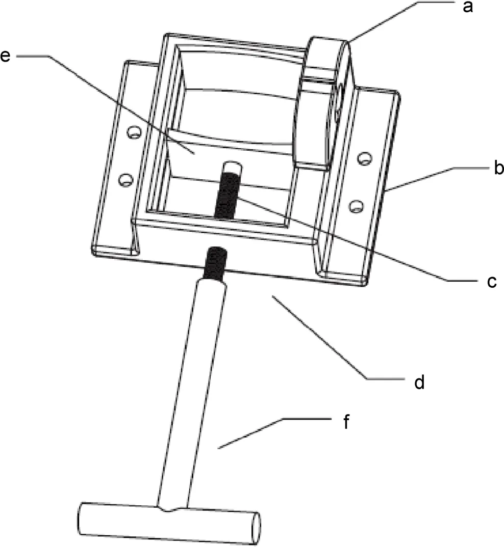
Figure 2 Top view of the fixture. a. Plastic diverter with memory function; b. skin‐fixation wings; c. check nut; d. base with circular arc and chamfering of the edge; e. removable stopper; f. rotating handle.
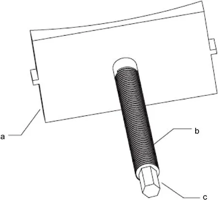
Figure 3 Schematic diagram of the removable stopper. a. Fixing baffle of the stopper; b. threaded rod; c. hexagonal head.

Figure 4 Schematic diagram of the rotating handle. a. Rotating handle; b. hexagonal internal head.
How is the device used? First, the drainage tube and surrounding skin are cleaned. Second, the base is fixed to the skin using the wings. The tube is then inserted through the hole and the rotating handle is used to move the limiting stopper towards the opposite side until the tube is pinched. This will prevent the tube from moving perpendicular to the skin. Finally, the tube is bent and inserted into the plastic diverter with memory function to prevent horizontal movementon the skin. Our experience shows that the new drainage tube device has a feasible design. In addition to being sewn or stapled to the skin, the removable stopper and plastic diverter with memory function work together to ensure that the drainage tube is fixed firmly (Figure 5). Therefore, the tube will not move either perpendicular or horizontal to the skin. In addition, the fixture can be used with drainage tubes with different specifications and materials. The limiting stopper can be moved easily, which enables local skin disinfection around the drainage tube. In conclusion, the device deserves clinical promotion.

Figure 5 Application of the drainage tube device. a. Drainage tube.
Conflict of interests
The authors have no financial interest to disclose regarding the article.
References
[1] Srinivasan VM, O’Neill BR, Jho D, Whiting DM, Oh MY. The history of external ventricular drainage. J Neurosurg 2014, 120(1): 228–236.
[2] Sun CR, Du HG, Yin LC, He M, Tian Y, Li HY. Choice for the removal of bloody cerebrospinal fluid in postcoiling aneurysmal subarachnoid hemorrhage: External ventricular drainage or lumbar drainage? Turk Neurosurg 2014, 24(5): 737–744.
[3] De Andrade AF, Paiva WS, Neville IS, Noleto GS, Junior AA, Sandon LHD, Bor-Seng-Shu E, Amorim RL, Teixeira MJ. Monoblock external ventricular drainage system in the treatment of patients with acute hydrocephalus: A pilot study. Med Sci Monit 2014, 20: 227–232.
[4] Kirmani AR, Sarmast AH, Bhat AR. Role of external ventricular drainage in the management of intraventricular hemorrhage; its complications and management. Surg Neurol Int 2015, 6: 188.
[5] Wang X, Dong Y, Qi XQ, Li YM, Huang CG, Hou LJ. Clinical review: Efficacy of antimicrobial-impregnated catheters in external ventricular drainage—A systematic review and meta-analysis. Crit Care 2013, 17(4): 234.
[6] Wiegand J, Hickson L, Merz TM. Indicators of external ventricular drainage-related infections—A retrospective observational study. Acta Neurochir 2016, 158(3): 595–601.
Call for papers
 Translational Neuroscience and Clinics2016年1期
- Translational Neuroscience and Clinics的其它文章
- Looking for positive viewpoints for Alzheimer’s caregivers: Case evaluation of a positive energy group
- Effects of the APOE ε4 allele on therapeutic response in Alzheimer’s disease
- Repairing skull defects in children with nano-hap/collagen composites: A clinical report of thirteen cases
- Effects of regional cerebral blood flow perfusion on learning and memory function and its molecular mechanism in rats
- An association between the location of white matter changes and the behavioral and psychological symptoms of dementia in Alzheimer’s disease patients
- Effects of aging on working memory performance and prefrontal cortex activity: A time‐resolved spectroscopy study